HeartLung.AI Marks Unprecedented Scientific Milestone with 10 Presentations at ESC Congress 2026
Unprecedented company milestone highlights AI advances in cardiovascular imaging, risk prediction, and preventive care

Press Release Disclaimer: This is a press release distributed through the XPR Media network. It has not been independently verified by our newsroom.
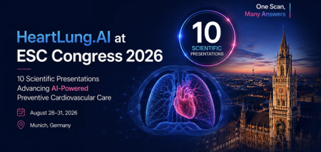

Unprecedented company milestone highlights AI advances in cardiovascular imaging, risk prediction, and preventive care from routine CT scans.
MUNICH , GERMANY, August 25, 2026 /EINPresswire.com/ — HeartLung.AI today announced that it will present an unprecedented 10 scientific presentations at ESC Congress 2026, taking place August 28–31 in Munich, Germany. The program represents the largest scientific showing at a single major cardiology congress in HeartLung.AI’s history and reflects years of collaboration among the company’s researchers, engineers, physicians and internationally recognized academic collaborators. HeartLung.AI has previously announced that it expects to be the only company at ESC Congress 2026 presenting this high number of accepted scientific studies.
The 10 studies span coronary artery calcium scoring, cardiac chamber volumetry, heart failure, atrial fibrillation, stroke, aortic stenosis, epicardial adipose tissue, cardiometabolic phenotyping, coronary plaque characterization and lung cancer risk prediction. Collectively, the research demonstrates a central theme behind HeartLung.AI’s work: extracting substantially more clinically meaningful information from CT scans that patients are already receiving.
“We are incredibly proud of our team and collaborators for reaching this milestone,” said Morteza Naghavi, MD, Founder and President of HeartLung.AI. “Having 10 scientific presentations at ESC Congress 2026 is unprecedented for our company and reflects the tremendous amount of work our researchers, engineers, physicians and academic collaborators have put into advancing image-guided prevention. What makes this program especially meaningful is the breadth of the science. These studies demonstrate how a single CT scan can potentially provide insights far beyond its original indication—from coronary disease and cardiac chamber abnormalities to future heart failure, atrial fibrillation, stroke, valvular disease and even lung cancer risk. I am extremely proud of our team and grateful to every investigator who contributed to this work.”
HeartLung.AI’s 10 Scientific Presentations at ESC Congress 2026
1. Comparison of AI-Enabled Cardiac Chambers Volumetry Based on Non-Gated Chest CT Scans with Echocardiography
Presenting Author: Seyed Reza Mirjalili, MD
This MESA study evaluated 1,675 participants who underwent both non-gated, non-contrast chest CT and echocardiography, examining whether AI-derived cardiac chamber measurements from routinely acquired chest CT can provide clinically meaningful information comparable with echocardiography for heart failure and atrial fibrillation assessment. The work supports the broader potential for opportunistic cardiovascular evaluation from millions of chest CT examinations already performed for other clinical reasons.
2. Long-Term Prediction of Incident Aortic Stenosis Using AI-CVD-Derived Phenotypes from Noncontrast Cardiac CT — The Multi-Ethnic Study of Atherosclerosis
Presenting Author: Zahra Heidari Meybodi, MD
The study analyzed 5,520 MESA participants over approximately two decades and assessed 25 AI-CVD-derived imaging phenotypes for their relationship with future aortic stenosis. Aortic valve calcification was the strongest individual predictor, while the broader AI-CVD model achieved a 10-year AUROC of 0.973 compared with 0.809 for the clinical model, demonstrating the potential to identify valvular disease risk years before clinical recognition.
3. Heart Failure and Atrial Fibrillation Prediction from Non-Gated Chest CT Using AI-Based Cardiac Chamber Volumetry — An AI-CVD Study within MESA
Presenting Author: Seyed Reza Mirjalili, MD
Among 2,053 MESA participants, AI-derived chamber measurements from routine non-gated chest CT demonstrated high agreement with measurements obtained from ECG-gated cardiac CT, including ICCs of 0.94 for left atrial volume, 0.95 for left ventricular volume and 0.96 for LV mass. Importantly, non-gated CT measurements were non-inferior to gated CT measurements for predicting future heart failure and atrial fibrillation, supporting a potentially scalable approach to opportunistic cardiac risk assessment.
4. Automated Epicardial Adipose Tissue Analysis Identifies Increased Non-Calcified Plaque Burden in Non-Obese Individuals with Low CAC Scores — An AI-CVD Study within the Miami Heart Study
Presenting Author: Hamed Zarei, MD
In 1,259 Miami Heart Study participants with paired CAC and CCTA scans, AI-derived epicardial adipose tissue measurements identified substantial differences in non-calcified coronary plaque burden that were not apparent from BMI or CAC score alone. Among participants with BMI below 29, non-calcified plaque burden increased approximately 93% per standard-deviation increase in epicardial fat, while low-CAC participants in the highest epicardial-fat quartile had approximately three times the median non-calcified plaque burden of those in the lowest quartile.
5. Agatston-2.0: AI-Derived Calcium Burden and Plaque Density Profiling Improve CHD Risk Stratification Within CAC Scores 1–99
First Author: Amir Azimi, MD
This study examined 1,542 participants from MESA and the Framingham Heart Study with a median 12.5 years of follow-up. Agatston-2.0 goes beyond the conventional calcium score by evaluating plaque density, distribution and related calcium characteristics. Within the traditionally broad CAC 1–99 category, 33% of participants were up-classified and accounted for 52% of 10-year CHD events, while 41% were down-classified to less than 5% 10-year risk. The approach produced a +34.8% net reclassification using information from the same CAC scan.
6. Cardiometabolic Phenotyping from Coronary Artery Calcium Scans Predicts Obstructive and High-Risk Plaques on Coronary CT Angiography — An AI-CVD Study within the Miami Heart Study
Using paired CAC and CCTA imaging from 1,259 asymptomatic Miami Heart Study participants, investigators evaluated whether comprehensive AI-CVD phenotyping could identify coronary disease characteristics beyond what is captured by traditional calcium scoring. The AI-CVD model achieved an AUC of 0.957 for obstructive stenosis compared with 0.881 using Agatston CAC alone, 0.789 versus 0.626 for high-risk plaque, and 0.771 versus 0.679 for non-calcified plaque. The findings demonstrate the potential for a routine CAC scan to serve as a much broader cardiometabolic imaging examination.
7. Agatston-2.0: A Next-Generation AI-Based Coronary Calcium Quantification Approach to Improve Risk Stratification Among Individuals with Zero Agatston Scores — Part I
Led by: Morteza Naghavi, MD
This study evaluated 3,965 individuals from MESA and the Framingham Heart Study who had a conventional CAC score of zero. Agatston-2.0 detected AI-derived coronary calcium in 21.7% of these individuals. Those with AI-CAC greater than zero demonstrated significantly higher long-term coronary heart disease risk and greater progression to conventional CAC positivity. Agatston-2.0 uses the same non-contrast CT scan without additional radiation while applying AI-based coronary segmentation, calibration, continuous voxel-level density weighting and spatial filtering to detect subtle calcification that conventional threshold-based scoring can miss.
8. Long-Term Lung Cancer Risk Prediction Using Sybil AI on Routine Coronary Artery Calcium Scans
This study evaluated baseline CAC scans from 5,726 MESA participants with follow-up extending up to 15 years. Using only imaging from the baseline CAC scan—and without smoking history or other clinical risk factors—Sybil AI identified imaging signals associated with future lung cancer risk. Predictive performance remained near 70% AUC through much of long-term follow-up and approximately 68% at 15 years, highlighting the possibility that cardiac CT examinations could contribute to lung cancer risk assessment without requiring another scan.
9. AI-Derived Left Atrial Volume Index, LA/RA and LA/LV Volume Ratios from Coronary Artery Calcium Scans Predict Long-Term Atrial Fibrillation and Stroke
Using MESA and Framingham Heart Study data with follow-up extending up to 17 years, investigators evaluated AI-derived left atrial volume index and cardiac chamber volume ratios from routine CAC scans for their association with future atrial fibrillation and stroke. The study demonstrates how automated chamber volumetry can extend a conventional coronary calcium examination beyond coronary disease assessment and provide additional information relevant to long-term cardiovascular risk.
10. Performance of Sybil AI for Lung Cancer Risk Assessment: A Head-to-Head Comparison of Cardiac vs. Lung CT Scans
Investigators evaluated Sybil AI on 4,486 cardiac CAC and chest CT scans from the Framingham Heart Study and MESA. Despite the more limited lung field of view available on cardiac CT, Sybil maintained an AUC above 0.80 for lung cancer risk prediction through six years. The findings suggest that CT scans originally obtained for cardiovascular prevention may also contain clinically meaningful information about future lung cancer risk.
A Multidisciplinary Scientific Collaboration
The breadth of the ESC Congress 2026 program reflects HeartLung.AI’s collaboration with internationally recognized investigators across cardiovascular medicine, radiology, preventive cardiology, imaging science and artificial intelligence.
Authors and collaborators across HeartLung.AI’s ESC 2026 scientific program include: Amir Azimi, MD; Kyle Atlas, MS; Chenyu Zhang, MS; Anthony P. Reeves, PhD; Seyed Reza Mirjalili, MD; Mohammadhossein MozafaryBazargany, MD; Amir Ghaffari Jolfayi, MD; Ali Hashemi; H. Mohammadi, MD; Hamed Ghoshouni, MD; Hamed Zarei, MD; Zahra Heidari Meybodi, MD; Zahi A. Fayad, PhD; David F. Yankelevitz, MD; Nathan D. Wong, PhD, MPH, FACC; Thomas Atlas, MD; Jakob Wasserthal, PhD; Oren Mechanic, MD, MPH, MBA; Rozemarijn Vliegenthart, MD, PhD; Claudia I. Henschke, PhD, MD; Andrea D. Branch, PhD; Jamal S. Rana, MD, PhD, FACC; Koen Nieman, MD, PhD; Jagat Narula, MD, PhD; Kim A. Williams Sr., MD; Prediman K. Shah, MD; Roxana Mehran, MD; Paolo Raggi, MD; David J. Maron, MD; Michael V. McConnell, MD, MSEE; Robert A. Kloner, MD, PhD; Matthew J. Budoff, MD; Arthur Agatston, MD, FACC; and Morteza Naghavi, MD.
“This achievement belongs to the entire team,” added Dr. Naghavi. “It is the product of rigorous scientific work, collaboration across institutions and a shared belief that the information already contained in medical images can be used much more effectively to identify disease earlier. We are honored to bring this body of work to ESC Congress 2026.”
From a Calcium Score to a Multidimensional Prevention Scan
Taken together, the 10 presentations demonstrate a broader evolution in CT-based prevention. Rather than treating a CT examination as a source of a single measurement, HeartLung.AI’s research program examines how artificial intelligence can extract multiple cardiovascular and multisystem biomarkers from the same existing images.
The studies range from refining the traditional Agatston calcium score with Agatston-2.0 to quantifying cardiac chambers, epicardial fat and cardiometabolic phenotypes, predicting aortic stenosis, heart failure, atrial fibrillation and stroke, and evaluating whether cardiac CT can contribute to long-term lung cancer risk assessment. The common objective is to obtain more actionable preventive information from imaging that has already been performed.
Full ESC Congress 2026 scientific presentations are available through the ESC 2026 section of HeartLung.AI’s Slide Presentations page: heartlung.ai/slide-presentations
About HeartLung.AI
HeartLung.AI is a health-tech company pioneering AI-driven preventive imaging for early detection of cardiovascular disease, lung cancer, COPD, osteoporosis, fatty liver disease, myosteatosis and other cardiometabolic conditions detectable on routine medical imaging. Its FDA-cleared flagship platform, AI-CVD, transforms eligible CT scans into comprehensive preventive health assessments by automatically quantifying coronary artery calcium, aortic and valvular calcification, cardiac chamber size, aorta and pulmonary artery size, epicardial and visceral fat, liver density, lung density, bone mineral density and muscle-fat composition. HeartLung requires no new hardware or local software installation. Hospitals and imaging centers can connect PACS to the HeartLung.AI cloud or manually upload scans and receive AI-generated DICOM and PDF reports.
Marlon Montes
HeartLung Technologies
+1 310-510-6004
email us here
Visit us on social media:
LinkedIn
YouTube
X
Legal Disclaimer:
EIN Presswire provides this news content “as is” without warranty of any kind. We do not accept any responsibility or liability
for the accuracy, content, images, videos, licenses, completeness, legality, or reliability of the information contained in this
article. If you have any complaints or copyright issues related to this article, kindly contact the author above.

Media gallery